
What an AI protein structure can tell us
A reader's guide to predicted coordinates, confidence scores, and the experimental conditions behind a protein's shape.

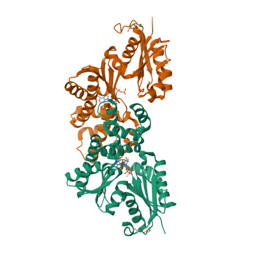
A molecular rendering of a protein structure presents a compelling visual narrative. Polypeptide chains wind through space as smooth ribbons, alpha helices coil into cylinders, beta sheets assemble into pleated ribbons, and coordinated cofactors nestle into deep binding pockets. Yet every structural diagram is fundamentally a visual abstraction of three-dimensional atomic coordinates deposited in databases such as the Protein Data Bank (PDB). Interpreting a structural model requires understanding the methodology that generated its coordinates, assessing quantitative confidence metrics, and contextualizing the model within the thermodynamic and biochemical conditions of the biological system.
Proteins are linear heteropolymers composed of twenty canonical L-amino acids joined by planar peptide bonds. Each amino acid residue comprises a common main-chain backbone () and a variable side chain () that imparts distinct physical characteristics, ranging from nonpolar hydrophobic residues (such as leucine and phenylalanine) to charged residues (such as aspartate and arginine). The spatial folding of a polypeptide chain is driven by hydrophobic collapse, hydrogen bonding networks, electrostatic interactions, and van der Waals packing, which position residues that are distant in primary sequence into intimate three-dimensional proximity.
In structural biology, molecular visualization software translates coordinates into stylized cartoon representations: ribbon trajectories follow backbone torsion angles (), tracing secondary structure while omitting explicit water molecules and mobile side chains. Experimental coordinates determined via X-ray crystallography reflect electron density maps averaged across billions of molecules within a crystal lattice. Cryo-electron microscopy (cryo-EM) reconstructs spatial densities from single-particle projections frozen in vitreous ice, while nuclear magnetic resonance (NMR) derives conformational ensembles from distance constraints in solution. Each experimental modality provides an empirical coordinate record conditioned by solvent pH, ionic strength, temperature, and crystal contact constraints.
Evolutionary co-variation and machine learning pipelines
Deep learning architectures for protein structure prediction, most notably AlphaFold2 (Jumper et al., 2021), fundamentally transformed structural biology by predicting 3D atomic coordinates directly from primary amino acid sequences. Rather than simulating physical folding pathways through molecular dynamics, AlphaFold2 relies on statistical patterns of evolutionary conservation and co-variation extracted from Multiple Sequence Alignments (MSAs).
When a mutation occurs at a residue position involved in a crucial tertiary contact, maintaining structural stability and biological function often requires a complementary mutation at the contacting residue across evolutionary timescales. Direct Coupling Analysis (DCA) and self-attention mechanisms detect these correlated mutations within large homologous sequence families, providing spatial proximity constraints.
AlphaFold2 processes these sequence alignments through an Evoformer module that iteratively exchanges representations between an MSA matrix and a pairwise residue-residue distance matrix via axial attention and triangular multiplicative updates. Subsequently, an Invariant Point Attention (IPA) Structure Module operates on local rigid-body frames, generating continuous Euclidean coordinates for all backbone and side-chain heavy atoms. In the 14th Critical Assessment of Structure Prediction (CASP14) in 2020, AlphaFold2 achieved unprecedented accuracy, predicting many single-domain proteins with backbone root-mean-square deviations (RMSD) below 1.5 angstroms compared to withheld experimental targets (Kryshtafovych et al., 2021).
Conformational ensembles versus static coordinate files
A fundamental principle of protein biophysics is that proteins in solution do not occupy a single static conformation. Instead, they populate dynamic thermodynamic ensembles distributed across complex free-energy landscapes.
A prime illustration is provided by the archaeal hexokinase from Sulfolobus tokodaii, investigated crystallographically by Nishimasu et al. (2007, pages 9923 to 9931). The unliganded apo enzyme crystallizes in an open conformation (PDB 2E2N), where two distinct structural domains are separated by a broad, solvent-accessible inter-domain cleft. Upon binding its physiological substrates (D-xylose, ADP, and magnesium ions), the two domains undergo a dramatic rigid-body hinge closure of approximately 14 degrees, engulfing the substrate within a closed active site (PDB 2E2Q).
Because AlphaFold2 was trained predominantly on static crystal structures deposited in the PDB, its primary output is a single coordinate configuration. For proteins possessing multiple functional states, an AI prediction typically corresponds to one favored state (often the apo form or whichever state dominates sequence homologues in the training corpus). It does not provide the complete thermodynamic partition function, activation barriers, or transition rates connecting conformational substates.
Local structural confidence: the pLDDT metric
To evaluate the local reliability of predicted coordinates, AlphaFold computes a per-residue confidence metric termed the predicted Local Distance Difference Test (pLDDT), spanning a continuous scale from 0 to 100.
The true Local Distance Difference Test (lDDT), formulated by Mariani et al. (2013), evaluates the local agreement of inter-atomic distances between a predicted model and an experimental reference structure without requiring global structural superposition:
where defines the local spherical inclusion radius around residue , denotes distance deviation tolerance thresholds (, , , and ), and is the indicator function. By evaluating conserved pairwise distances within a 15-angstrom neighborhood, lDDT is robust against domain hinge motions that degrade global RMSD metrics.
AlphaFold2 trains a dedicated projection head to predict the expected lDDT score for each atom directly from the network representations, producing the pLDDT score. Interpreting pLDDT requires applying standard empirical thresholds established by EMBL-EBI and the AlphaFold team:
- (Very High Confidence): Backbone and side-chain rotamers exhibit high precision, often rivaling experimental crystal structures below 2-angstrom resolution. These regions are suitable for analyzing catalytic mechanisms and atomic contacts.
- (Confident): The polypeptide backbone trace and secondary structure are modeled reliably, though individual surface side-chain rotamers may vary.
- (Low Confidence): The overall fold or loop topology may be plausible, but specific coordinates and bond geometries are uncertain.
- (Very Low Confidence): Frequently indicates intrinsically disordered regions (IDRs) that lack a fixed equilibrium tertiary structure in isolation (Tunyasuvunakool et al., 2021). When visualized in 3D, these segments often appear as extended ribbon loops resembling cooked spaghetti, reflecting the absence of stable internal contacts rather than an experimental coordinate artifact.
Global domain placement: the Predicted Aligned Error matrix
While pLDDT quantifies the local accuracy of the immediate atomic neighborhood surrounding a single residue, it provides no information about the relative spatial orientation between distant domains. To evaluate global quaternary and inter-domain arrangement, AlphaFold computes the Predicted Aligned Error (PAE) matrix.
Formally, let denote the local Euclidean reference frame of residue , defined by the coordinates of its backbone atoms (, , ). When the predicted model is rigidly superposed onto the true physical structure by aligning their local frames at residue , the Euclidean position error of residue is:
AlphaFold predicts a probability distribution over the continuous error discretized into 64 distance bins up to 32 angstroms. The expected value under this distribution defines the predicted aligned error:
Because the alignment is anchored to the local orientation of residue , the metric is asymmetric: . A low indicates that aligning the structure on residue produces a precise, low-variance position for residue .
In the 2D PAE plot, rows represent residue and columns represent reference residue . For a multi-domain protein, well-folded individual domains appear as square blocks along the diagonal with uniform low error (typically 0 to 4 angstroms). If the off-diagonal rectangular regions comparing residues between domain 1 and domain 2 show high expected error (such as 15 to 30 angstroms), the model possesses high confidence in each domain's internal fold, but no confidence in their relative orientation. In such cases, the domains are likely connected by a flexible linker, and treating the visual domain-domain interface as a fixed physiological interaction would be an error.
From single-chain folds to biomolecular complexes: AlphaFold 3
The original AlphaFold2 architecture was specialized for single polypeptide chains and subsequently extended to multimeric protein complexes (AlphaFold-Multimer). However, biological function in vivo frequently depends on non-protein components: divalent metal cofactors (), nucleic acid scaffolds (DNA, RNA), post-translational modifications (phosphorylation, glycosylation), and small-molecule metabolites or pharmacological drugs.
AlphaFold 3 (Abramson et al., 2024) introduced a major architectural paradigm shift. It replaced the Evoformer with a streamlined Pairformer that operates directly on pair representations without full MSA attention blocks. Crucially, AlphaFold 3 removed the invariant rigid-body frame representation of the Structure Module, replacing it with a generative 3D Diffusion Module. The diffusion network operates directly on raw 3D Cartesian coordinates of all heavy atoms, iteratively denoising random Gaussian coordinate clouds into folded biomolecular assemblies.
This unified generative formulation allows AlphaFold 3 to predict joint complexes comprising proteins, nucleic acids, ligands, and ions simultaneously. However, predicting small-molecule binding poses remains challenging: while global complex topology is frequently accurate, precise atomic coordinating bonds and catalytic side-chain orientations still require empirical validation through cryo-EM or high-resolution crystallographic co-complex structures.
A reading protocol for predicted structural models
To critically interrogate a predicted protein structure from databases such as AlphaFold DB (Varadi et al., 2022) or research publications, researchers should follow a structured diagnostic protocol:
- Verify Sequence Context and Boundaries: Confirm the exact UniProt accession, construct boundaries, and multiple sequence alignment depth. Stretches with shallow alignment depth ( effective sequences) often exhibit lower predictive accuracy.
- Inspect Residue-Level pLDDT Scores: Examine the local confidence profile along the specific functional region under study (such as an enzyme active site or epitope). Do not rely on whole-chain mean pLDDT, which can obscure low-confidence loops embedded in rigid domains.
- Analyze the PAE Matrix for Multi-Domain Proteins: Check whether the off-diagonal blocks between domains exhibit low expected error (). High off-diagonal error indicates flexible inter-domain hinging, warning against over-interpreting relative domain orientations.
- Contextualize Biochemical State and Conformational Multiplicity: Determine whether the biological question requires an open apo state or a ligand-bound closed holo conformation. Check for missing cofactors, prosthetic groups, or oligomeric partners that govern physiological folding.
Executing this protocol ensures that computational coordinate predictions are evaluated with the rigorous methodological discipline required of modern molecular biophysics.
References
- Abramson, J., Adler, J., Dunger, J., Evans, R., Green, T., Pritzel, A., Ronneberger, O., Willmore, L., Yim, A. K., Ding, J., Qiu, L., Wu, Z., Lovas, S., Meyer, T., Wickline, T., Wang, Z., ... Jumper, J. M. (2024). Accurate structure prediction of biomolecular interactions with AlphaFold 3. Nature, 630(8016), 493-500. https://doi.org/10.1038/s41586-024-07487-w
- Jumper, J., Evans, R., Pritzel, A., Green, T., Figurnov, M., Ronneberger, O., Tunyasuvunakool, K., Bates, R., Žídek, A., Potapenko, A., Bridgland, A., Meyer, C., Kohl, S. A. A., Ballard, A. J., Cowie, A., ... Hassabis, D. (2021). Highly accurate protein structure prediction with AlphaFold. Nature, 596(7873), 583-589. https://doi.org/10.1038/s41586-021-03819-2
- Kryshtafovych, A., Schwede, T., Topf, M., Fidelis, K., & Moult, J. (2021). Critical assessment of methods of protein structure prediction (CASP): Round XIV. Proteins: Structure, Function, and Bioinformatics, 89(12), 1607-1617. https://doi.org/10.1002/prot.26237
- Mariani, V., Biasini, M., Barbato, A., & Schwede, T. (2013). lDDT: a local superposition-free score for comparing protein structures and models using distance difference tests. Bioinformatics, 29(21), 2722-2728. https://doi.org/10.1093/bioinformatics/btt473
- Nishimasu, H., Fushinobu, S., Shoun, H., & Wakagi, T. (2007). Crystal structures of an ATP-dependent hexokinase with broad substrate specificity from the hyperthermophilic archaeon Sulfolobus tokodaii. Journal of Biological Chemistry, 282(13), pages 9923 to 9931. https://doi.org/10.1074/jbc.M610678200
- Tunyasuvunakool, K., Adler, J., Wu, Z., Green, T., Zielinski, M., Žídek, A., Bridgland, A., Cowie, A., Meyer, C., Laydon, A., Velankar, S., Kleywegt, G. J., Bateman, A., Evans, R., Pritzel, A., ... Hassabis, D. (2021). Highly accurate protein structure prediction for the human proteome. Nature, 596(7873), 590-596. https://doi.org/10.1038/s41586-021-03828-1
- Varadi, M., Anyango, S., Deshpande, M., Nair, S., Natassia, C., Yordanova, G., Yuan, D., Stroe, O., Wood, G., Laydon, A., Žídek, A., Green, T., Tunyasuvunakool, K., Petersen, S., Jumper, J., ... Velankar, S. (2022). AlphaFold Protein Structure Database: massively expanding the structural coverage of protein-sequence space with high-accuracy models. Nucleic Acids Research, 50(D1), D439-D444. https://doi.org/10.1093/nar/gkab1061